Recent studies with silver nanoparticles (AgNPs) and the history of silver metal as a broad-spectrum bactericidal and virucidal agent, places silver as one of the future biocidal candidates in the field of nanomedicine to eliminate bacteria and viruses, especially multidrug resistant ones. In this review, we have described the various morphologies of AgNPs and correlated the enhanced bactericidal activity with their prominent {111} facets. In addition to prioritizing the characterization we have also discussed the importance of quantifying AgNPs and silver ion content (Ag+) and their different mechanisms at the chemical, biological, pharmacological, and toxicological levels. The mechanism of action of AgNPs against various bacteria and viruses including the SARS-CoV-2 was analyzed in order to understand its effectiveness as an antimicrobial agent with therapeutic efficacy and low toxicity. Further, there is the need to characterize AgNPs and quantify the content of free Ag+ for the implementation of new systematic studies of this promising agent in nanomedicine and in clinical practice.
PMCID: PMC7486059PMID: 32958250
https://pubmed.ncbi.nlm.nih.gov/22024958/
The elemental metal, Silver (Ag) has broad spectrum antimicrobial action against various bacteria, fungi and viruses. Due to their versatility, Ag nanoparticles (AgNP) have currently found their way as a microbicide for biological surfaces in various forms such as wound dressings, medical devices, deodorant sprays and fabrics. Several studies have demonstrated the potent antiviral action of AgNPs against various human pathogenic viruses such as Respiratory syncytial virus (RSV), Influenza virus, Norovirus, Hepatitis B virus (HBV) and Human immunodeficiency virus (HIV) [1]. In addition to these viruses, since Ag has been demonstrated to kill SARS-CoV, we hypothesized the strong possibility of AgNPs to inhibit SARS-CoV-2 [2,3]. Till date there are no studies directly demonstrating the effect of AgNPs on SARS-CoV-2. We tested colloidal silver (cAg), plain elemental Ag nanoparticles of different diameters (AgNPn) and polyvinylpyrrolidone capped 10 nm silver nanoparticles (PVP–AgNP10) against SARS-CoV-2 to find the most effective size and concentration of Ag that could inhibit SARS-CoV-2. We propose that AgNPs could be used on inanimate and non-biological surfaces to efficiently control the ongoing COVID-19 pandemic while simultaneously exercising care not to abuse it.
4.2. Silver inhibits extracellular viruses by preventing viral entry
We next performed the VPrA, cell post-treatment assay (CPoA) and the cell pre-treatment assay (CPrA) on SARS-CoV-2 using PVP-AgNP10 in VeroE6/TMPRSS2 to observe the effect of Ag on extracellular and intracellular viruses (Fig. 3 A). VPrA showed effective inhibition of extracellular free virions characterized by both the reduction of cell death and also a steep fall in the viral load to negligible levels (Fig. 3B and C). We further performed the CPoA to detect the ability of Ag to suppress virus in already infected cells. In this experimental design, VeroE6/TMPRSS2 cells were allowed to be infected with SARS-CoV-2 (MOI 0.05) for2 hours after which the extracellular viruses were washed and then the infected cells were treated with 2 ppm of PVP-AgNP10. We observed significant protection of infected cells and suppression of viral load with PVP-AgNP10 (Fig. 3B and C). Additionally we performed the CPrA to assess the ability of silver pre-treated cells to resist viral infection. VeroE6/TMPRSS2 cells were incubated with 2 ppm PVP-AgNP10 for 3 h after which the cells were washed to remove unbound silver followed by infection with SARS-CoV-2 (MOI 0.05). At the end of 48 h, the virus was found only to be partially inhibited (Fig. 3C).
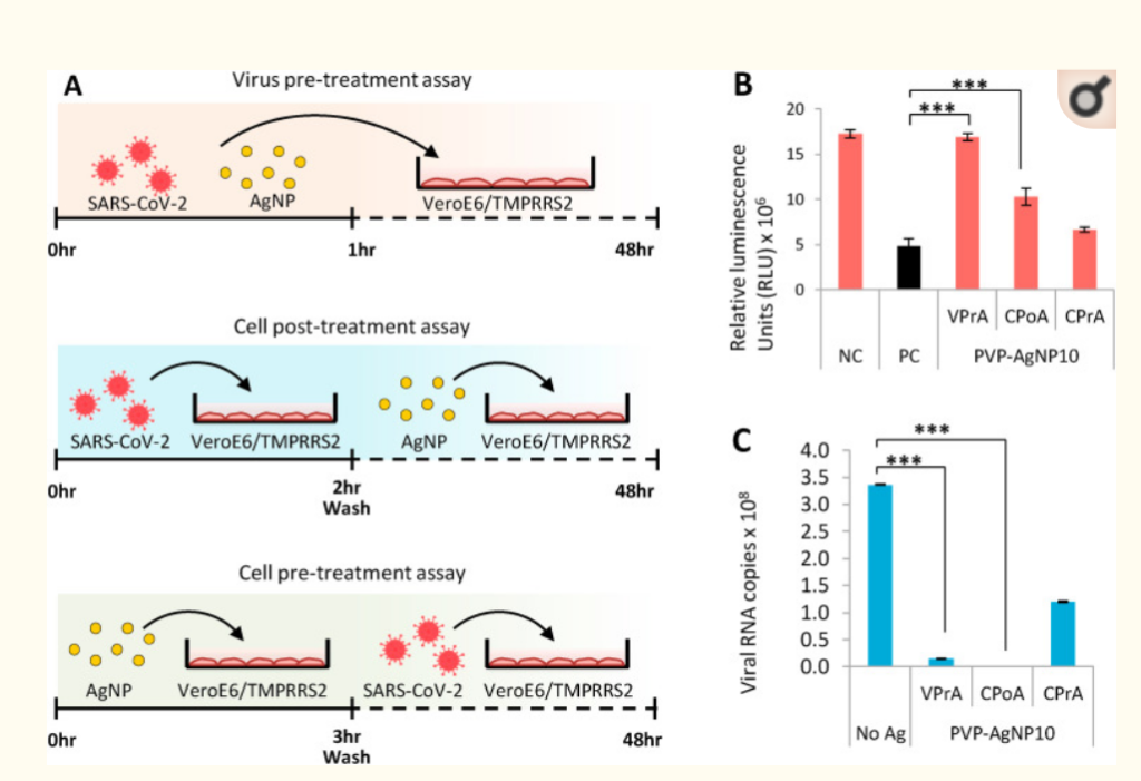
Silver nanoparticles effectively inhibit extracellular SARS-CoV-2.3A: Schematic representation of virus pre-treatment assay (top panel), cell post-treatment assay (central panel) and cell pre-treatment assay (bottom panel). 3B: Performance of PVP coated 10 nm Silver nanoparticles in the three study designs with respect to rescue of cells from SARS-CoV-2 infection. Error bars obtained from triplicate testing. p value ≤ 0.005 (∗∗∗). 2C: Performance of PVP coated 10 nm Silver nanoparticles in the three study designs with respect to reduction of SARS-CoV-2 replication. Error bars obtained from triplicate testing. p value ≤ 0.001 (∗∗∗).
We confirmed the size dependent antiviral effect of PVP- AgNP10 using the immunofluorescence analysis carried on VPrA experimental model; SARS-CoV-2 infection was effectively averted by AgNP10 and not by AgNP100 (Fig. 4 A). The plaque assay revealed that silver achieved complete inhibition of 0.05 MOI which is one log10 fold more than virus control. Partial inhibition was observed with higher viral loads starting from 0.5 MOI (Fig. 4B). To assess the role of AgNPs in viral entry, we performed the luciferase based pseudovirus entry assay. PVP-AgNP10 potently inhibited pseudoviral entry characterized by significant reduction of luciferase activity similar to that of the neutralizing antibody used as control (Fig. 4C).
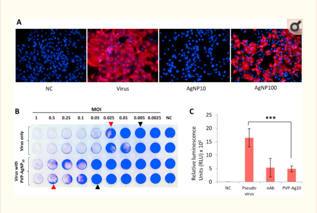
Characteristics of PVP coated 10 nm Silver nanoparticles in SARS-CoV-2 infection. 4A: Immunofluorescence imaging comparing the effect of 10 nm and 100 nm Silver nanoparticles against SARS-CoV-2 infection in VeroE6/TMPRSS2 cells. Cell nuclei (blue) and SARS-CoV-2 nucleocapsid protein in cytoplasm (red). NC – Negative control. 4B: PVP coated 10 nm Silver nanoparticles protect VeroE6/TMPRSS2 cells from SARS-CoV-2 infection mediated cell death. Crystal violet staining reveals partial protection with visible plaques (red arrowheads) and complete protection with absence of plaques (black arrowheads). 4C: Pseudovirus entry assay. PVP coated 10 nm Silver nanoparticles inhibit entry of pseudovirus in VeroE6/TMPRSS2 cells. NC – Negative control, nAb – Neutralizing antibody. (For interpretation of the references to colour in this figure legend, the reader is referred to the Web version of this article.)
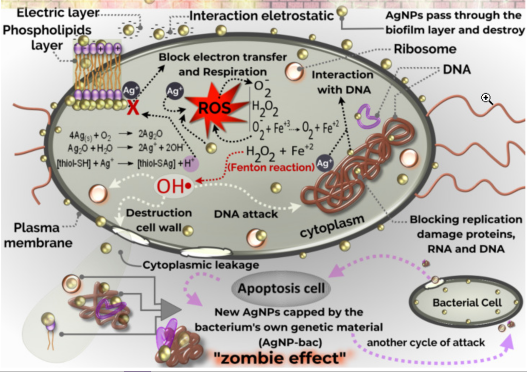
Ag is long known for its antimicrobial effect and the antiviral property of AgNPs is being extensively researched with renewed interest in the recent past [1]. The exact mechanism by which AgNPs exert its killing effect on viruses is still obscure. However, it has been consistently observed that AgNPs interact with the structural proteins on the surface of extracellular viruses to inhibit infection in the early phase, by either preventing viral attachment or entry, or by damaging the surface proteins to affect the structural integrity of virions [11,12]. In the current study, we have obtained similar findings in the VPrA where AgNPs effectively inhibits extracellular SARS-CoV-2 to protect the target cells from infection and the pseudovirus entry assay revealed that AgNPs interfere with viral entry.
AgNPs have been shown to preferentially bind to viral surface proteins rich in sulfhydryl groups and cleave the disulfide bonds to destabilize the protein, thereby affecting viral infectivity [11,13]. Studies on HIV have shown that AgNPs associate to the disulfide bonds that are in close proximity to the CD4 binding domain of the gp120 surface protein [11]. Hati and Bhattacharyya have demonstrated the importance disulfide bonds in binding of SARS-CoV-2 spike protein with the angiotensin converting enzyme-2 (ACE2) receptor and the disruption of which lead to impaired viral binding to the receptor [14]. Considering the mechanism of action of AgNPs shown by other authors, it can be presumed that AgNPs exert their antiviral effect on SARS-CoV-2 by disrupting the disulfide bonds on the spike protein and ACE2 receptors. Further studies are being conducted to find the antiviral mechanism of AgNPs on SARS-CoV-2 and elucidate it in detail subsequently.
AgNPs have also been claimed to possess intracellular antiviral action by interacting with viral nucleic acids [15]. We observed a partial antiviral effect in CPrA, as there was some amount of reduction in the viral load in cells pre-treated with PVP-AgNP10. While the reason for this effect is not known at present, it is possibly explained to be either due to the destruction of disulfide bridges on ACE2 receptor or due to a true intracellular mechanism (there by inhibiting serial viral infection of newly produced virus from infected cells to uninfected cells). Also, since Ag binds non-specifically to proteins, their use as antiviral agents might also cause some cellular dysfunction. Further studies are required to more precisely explain the holistic effect of Ag in vivo.
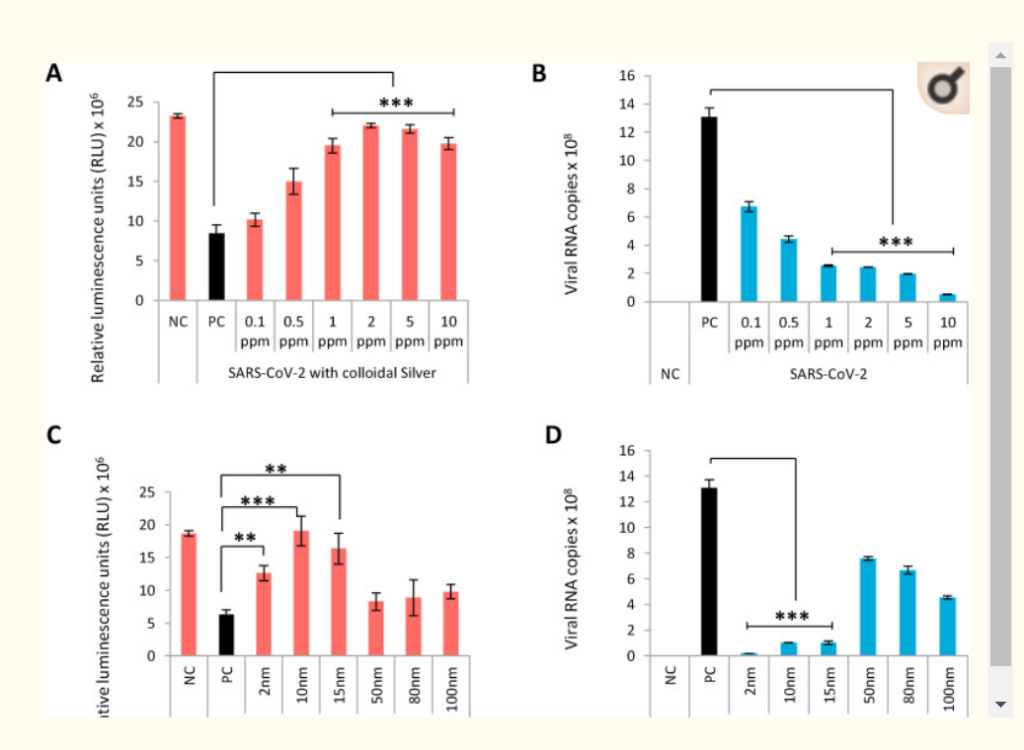
Several studies have reiterated the size dependent antiviral effect of AgNPs with particles around 10 nm diameter being most effective [1]. This has been attributed the higher stability of interaction to the viral protein achieved by 10 nm particles which is not capable by larger particles [11]. Consistent with this, we also observed anti-SARS-CoV-2 activity only with AgNPs of diameters ranging from 2 to 15 nm. Our immunofluorescence study corroborated the above phenomenon, as we observed that PVP-AgNP10 completely inhibited SARS-CoV-2 but AgNP100 did not.
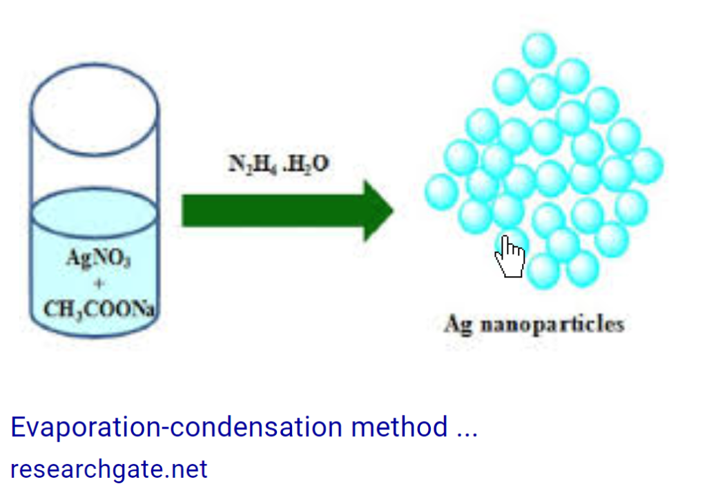
AgNPs can be generated by several methods and can contain reducing agents and capping agents along with the metal particles [16]. Coated or capped AgNPs are found to be more advantageous than plain AgNPs as coating increases stability, decreases agglomeration and reduces cytotoxicity of AgNPs [17]. Among the coated AgNPs, PVP capped nanoparticles are widely studied for biological use. It has been observed that PVP coating of AgNPs does not hinder their antiviral activity while other coating agents do [18]. PVP-AgNP10 has been demonstrated to possess excellent antiviral activity against enveloped viruses such as RSV and HIV [11,19]. This was the rationale to select PVP-AgNP10 for the study and we have demonstrated the robust antiviral effect of PVP-AgNP10 against SARS-CoV-2.
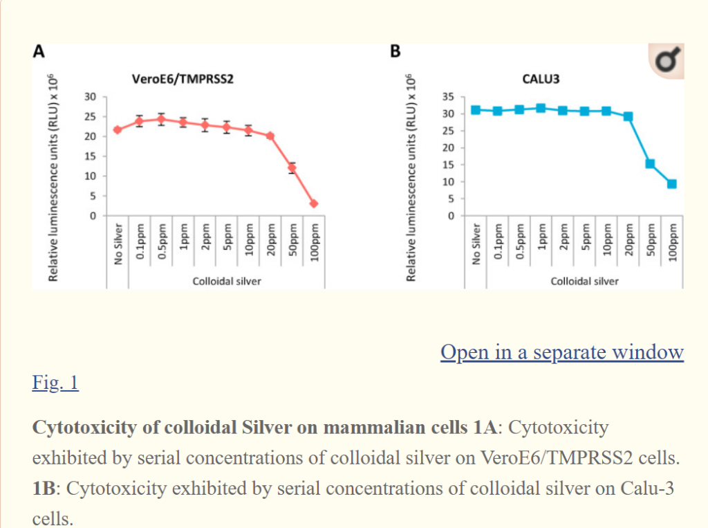
Antiviral effect of AgNPs is also concentration dependent. Most studies have observed the antiviral efficacy of AgNPs at concentrations ranging between 10 and 100 ppm [1]. However, 0.5 ppm cAg has been shown effective in inhibiting Influenza virus and is the least concentration that has been reported to show antiviral activity [20]. In the current study, we observed naked AgNPs to inhibit SARS-CoV-2 at concentrations ranging between 1 and 10 ppm and become cytotoxic to mammalian cells from 20 ppm and above.
Cytotoxicity of AgNPs to mammalian cells depends on the cell type and also the type of AgNPs. Mehrbod et al. have observed cytotoxicity in Madin-Darby Canine Kidney (MDCK) cells with naked cAg particles at concentrations higher than 0.5 ppm [20]. Naked AgNPs with NaBH4 reducing agent were found to induce apoptosis in colon adenocarcinoma cells at 11 ppm, while Citrate-stabilized naked AgNPs have been observed to exhibit cytotoxicity at concentrations higher than 30 ppm [21,22]. In this regard, PVP coated AgNPs have been demonstrated to be the least cytotoxic with no demonstrable cytotoxicity even at 50 ppm in human alveolar basal epithelial cells [19]. Smaller particles have a higher toxic potential due to the greater surface area of interaction with the bound protein [23]. We observed this effect as AgNP2 showed cytotoxicity even at 2 ppm while none of the bigger particles were cytotoxic at this concentration. Therefore, care should be exercised when Ag is used on biological surfaces.
Various ingestible and inhalable formulations of Ag are being marketed as cure for COVID-19, which available to purchase over the counter. The cytotoxic potential of these formulations should be considered before personal use. Also, Ag is a very broad spectrum microbicide. Illicit use of Ag might create an imbalance in the commensal microbiota leading to unforeseen consequences [24]. AgNPs can be used on a variety of inanimate surfaces to combat the ongoing COVID-19 pandemic [3]. Ag coated masks have been found to be effective in inhibiting SARS-CoV-2 and could potentially be effective when applied on the air filters of air conditioners and medical devices [25]. AgNP incorporated polycotton fabrics have been proven to inhibit SARS-CoV-2 [26]. Ag based sanitizers and disinfectants are also being used for disinfection of hands and inanimate surfaces respectively [27]. However, the effect of AgNPs on influencing the microbial life when released in the environment is unknown [16]. A proper disposal protocol should be developed for Ag containing products to avoid causing untoward imbalances in the environmental microbial pattern when discarded after use.